FIG. 7.
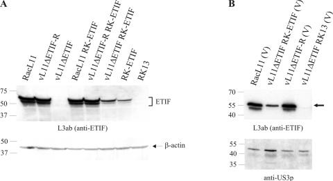
(A) Detection of ETIF in lysates of RK13 and RK-ETIF cells infected with the indicated viruses 18 h p.i. Lysates were separated by SDS-12% PAGE, transferred to a polyvinylidene difluoride membrane, and incubated with either anti-ETIF MAb L3ab or a control anti-β-actin MAb. (B) Purified virions of RacL11, vL11ΔETIF propagated on RK-ETIF cells, vL11ΔETIF-R, and vL11ΔETIF grown on RK13 cells were incubated with either anti-ETIF MAb L3ab or control anti-US3p polyclonal antibody. Only the full-length 60-kDa form of ETIF is indicated by a solid arrowhead. The sizes of the Precision Plus Protein (Bio-Rad) marker are given in thousands.

