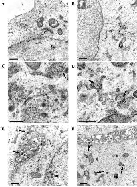
FIG. 9.
Electron micrographs of RK13 (A to D) or RK-ETIF (E and F) cells infected with vL11ΔETIF at 16 h p.i. (A) Overview of a section of an infected cell containing nucleocapsids in the nucleus and cytoplasmic nucleocapsids in the cytoplasm. No completely enveloped virions could be detected in the cytoplasm or extracellular space. (B) Nonenveloped cytoplasmic nucleocapsids released from infected-cell nuclei accumulated in the neighborhood of stacks of cytoplasmic membranes. (C) A cytoplasmic particle is budding into a presumably TGN originated membrane as indicated by an arrow. (D) Higher magnification demonstrated fuzzy material around cytoplasmic nucleocapids while budding into TGN membranes (arrows). (E) Infection of RK-ETIF with vL11ΔETIF at 16 h p.i. revealed nucleocapsids in the nucleus of infected cells, some which are adjacent to the nuclear envelope (arrowhead) and many fully enveloped particles in the extracellular space (arrows). (F) Secondary enveloped particles in the cytoplasm and extracellular virions are indicated by arrows. Bars, 500 nm (A to F).

