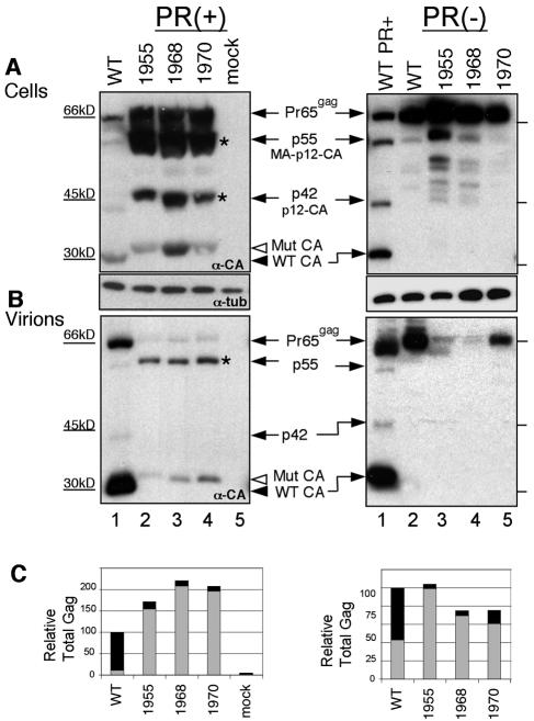
FIG. 2.
Gag proteins of release-defective mutants. (A) Viral protein expression in cells transfected with proviral DNA. Western blot analysis using anti-CA polyclonal serum detected CA and other Gag intermediates 48 h after transfection. The arrow indicates the position of the Gag precursor (Pr65gag) and intermediates. Open arrowhead, mutant CA protein; closed arrowhead, WT CA protein. Asterisks mark intermediates of Gag processing. The lower blot in panel A is the same blot and was probed with antitubulin antibody. Mutants in the context of an inactive viral protease are shown in the right panel. (B) Virions released into the medium following transfection with proviral DNA were concentrated and lysed, and the proteins were analyzed by Western blotting using anti-CA antibodies. (C) Particle release was defective for mutants 1955, 1968, and 1970. Gag proteins were quantified from blots. Gray bars represent intracellular Gag protein (from panel A, using lower exposures to ensure that signals were in the linear range for film), and black bars denote Gag released in pelleted virions (from panel B). The total amount of Gag protein in wild-type virus was set to 100%.

