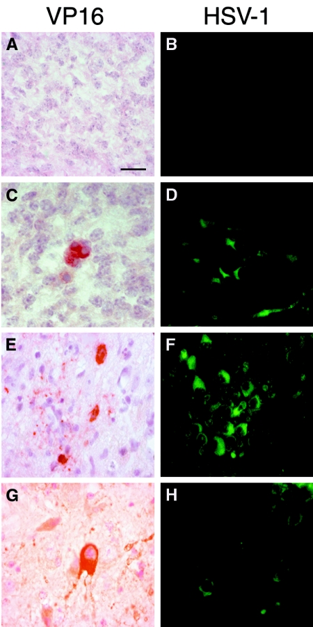
FIG. 6.
Immunohistochemical detection of HSV-1 antigens in the encephalon of fetuses and neonates. A group of seven female mice were infected intraperitoneally with 106 PFU of HSV-1 (KOS strain) and mated at 37 days postinfection with mock-infected male mice. From these, 15 fetuses were obtained for analysis on day 18 of gestation, plus another 30 neonates which were analyzed at 1 day postdelivery. Their organs were embedded in paraffin wax and serially sectioned. Immunodetection was performed by using two specific antibodies: anti-VP16 (against the tegument viral protein, revealed by a red chromogenic substrate) and anti-HSV-1 (which recognizes viral glycoproteins, detected by indirect immunofluorescence) as described in Materials and Methods. To rule out nonspecific viral staining, several controls were performed. The sections of fetal encephalon obtained from mock-infected female mothers showed no reaction to VP16 (A) or HSV-1 (B) antigens. (C) VP16 specific staining was observed, however, in some neurons of the cortex of fetal encephalons obtained from infected female mice. (D) Similarly, HSV-1 glycoprotein signals were detected by immunofluorescence in several neurons of the fetal encephalon from infected mothers at a similar location as described above, although more infected cells were visible. (E and F) Coronal sections of female neonate brains from infected mothers showed specific VP16 staining in a few neurons of the midbrain region (E), while HSV-1 glycoprotein signals were detected in a higher number of neurons (F). (G and H) Sections of male neonate encephalons from infected mothers showed specific immunoreactions with VP16 in the cytoplasm of neurons from the midbrain region (G), while HSV-1 immunoexpression was detected more strongly in mesencephalic neurons (more weakly in male mice) (H). Bar, 20 μm.

