FIG. 7.
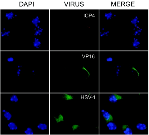
HSV-1 immunolocation in mouse fetal hippocampal cells from latently infected mothers. Female mice were infected intraperitoneally with 106 PFU of HSV-1 (KOS strain) and mated at 37 days postinfection with mock-infected male mice. Hippocampal cells were then obtained from fetuses at 18 days of gestation, plated, and after 1 day in vitro were analyzed by using several antibodies. Immunofluorescence analysis shows discrete dots marked with the anti-ICP4 antibody (middle top; red) and perinuclear signals stained with anti-VP16 (middle center; green). Cytoplasmic immunodetection was demonstrated by using an anti-HSV-1 marker (middle bottom; green). DAPI images of chromatin staining are shown on the left, and merge images are shown on the right.

