FIG. 2.
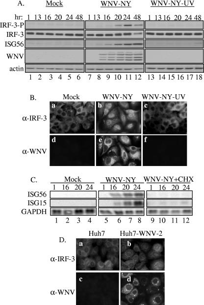
WNV-NY replication is required for activation of IRF-3. (A and B) Examination of IRF-3 activation in response to UV-inactivated WNV-NY. A549 cells were mock infected, infected with WNV-NY (MOI, 5), or exposed to UV-inactivated virus at a concentration equivalent to an MOI of 5. (A) Cell lysates were recovered at the indicated times and subjected to immunoblot analysis. Phosphorylation of IRF-3 was detected using an antibody specific for the phosphoserine 396 isoform of IRF-3 (IRF-3-P). Steady-state protein levels of total IRF-3, ISG56, WNV, and actin were also examined. (B) IRF-3 localization in mock-infected (a and d), WNV-NY-infected (b and e), or UV-inactivated WNV-NY- (c and f) treated A549 cells was detected by IFA. IRF-3 was detected using an IRF-3 polyclonal antiserum and an Alexa 488-conjugated secondary antibody (a, b, and c). WNV protein expression (d, e, and f) was detected using a mouse polyclonal anti-WNV antibody and rhodamine-conjugated secondary antibody. (C) Effect of cycloheximide on WNV-NY-induced expression of IRF-3 target genes. A549 cells were infected with WNV-NY (MOI, 1) in the presence or absence of cycloheximide (50 μg/ml). Induction of ISG15 and ISG56 was assessed by Northern blot analysis of total RNA harvested at the indicated times postinfection. Levels of GAPDH expression were also assessed to control for loading. (D) Activation of IRF-3 in cells harboring the WNV replicon. Cellular localization of IRF-3 (a and b) and WNV protein expression (c and d) were examined in parental Huh7 (a and c) and Huh7-WNV-2 replicon (b and d) cell lines by IFA.

