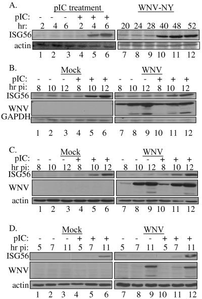
FIG. 5.
TLR3-mediated activation of IRF-3. (A) 293T-pCDNA-TL3-YFP cells were treated with pIC or infected with WNV-NY at an MOI of 5. Whole-cell lysates were recovered at the indicated times, and steady-state levels of ISG56 expression were examined by Western blotting. Blots were stripped and reprobed for actin to control for loading. (B) pIC was added to the culture supernatants of WNV-NY-infected (MOI, 3.6) 293T-pCDNA-TL3-YFP cells at 6 h postinfection. (C) U-2 OS/NS3/4A cells were infected with WNV-NY (MOI, 3) for 4 h prior to the addition of pIC to the culture medium. (D) PH5CH8 cells infected with WNV-NY (MOI, 1) were treated with pIC at 3 h postinfection. In panels B, C, and D, whole-cell lysates collected at the indicated times postinfection were analyzed for steady-state levels of ISG56 by immunoblotting. Blots were stripped and reprobed for WNV protein to assess viral replication and GAPDH or actin to control for loading.

