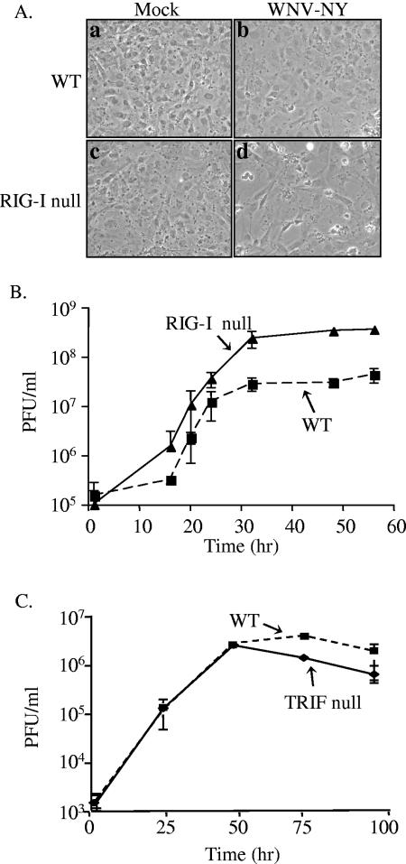
FIG. 8.
Replication of WNV-NY in WT, RIG-I null, and TRIF null MEFs. (A) Virus-induced CPE in WT and RIG-I null MEFs. Mock-infected (a and c) and WNV-NY-infected (b and d) cultures of WT (a and b) and RIG-I null (c and d) MEFs were visualized at 56 h postinfection using a Zeiss light microscope, and images were captured with a digital camera. (B) Infectious particle production by WNV-NY-infected WT and RIG-I null MEFs. Culture medium was removed from infected MEFs and cleared of cell debris by low-spin centrifugation. The presence of infectious virus particles was determined as PFU per milliliter by titrating supernatants on Vero cells in duplicate. The average of three independent experiments is shown. Solid line, RIG-I null; broken line, WT MEFs. (C) Infectious particle production by WNV-NY-infected TRIF null MEFs. Titers for supernatants removed from WNV-NY-infected WT and TRIF null MEFS were determined on Vero cells in duplicate. The average of three independent experiments is shown. Solid line, TRIF null; broken line, WT MEFs.

