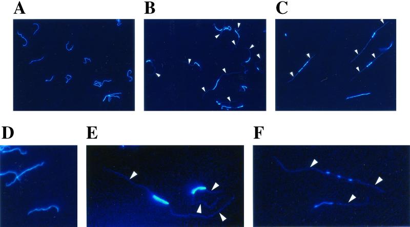
FIG. 5.
Fluorescence microscopy of L. biflexa cells stained with DAPI. Cells of exponential-phase cultures of L. biflexa wild-type strain (A and D) and L. biflexa recA mutant (B, C, E, and F). Arrows point to a faint fluorescence signal in the cellular space of the spirochetes. Pictures from the same groups (top panel: A, B, and C; bottom panel: D, E, and F) are enlarged to the same extent. All images are at the same magnification (original magnification, ×600).

