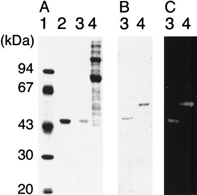
FIG. 3.
Expression of AgaA in C. josui and E. coli. The gel was stained with Coomassie brilliant blue (A) or stained for α-galactosidase activity (C). AgaA proteins were detected with a polyclonal mouse antiserum raised against truncated AgaA by Western blot analysis (B). Lane 1, protein mass standards; lane 2, truncated AgaA purified from a recombinant E. coli strain (1 μg); lanes 3, truncated AgaA purified from a recombinant E. coli strain (0.1 μg); lanes 4, cellulosomal proteins of C. josui.

