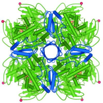
FIG. 6.
Tertiary structure of RubisCO, showing the position of the six-amino-acid insert in the Nitrosospira sp. large subunit. The L8S8 R. eutropha holoenzyme is shown, based on the structure of the unactivated enzyme, as determined by X-ray crystallography (23). Large subunits are green, and small subunits are blue. Phosphate ions bound in the active site are yellow. The C-alpha atom of Asn95 (corresponding to Asn91 in the Nitrosospira sp. isolate 40KI sequence) is represented as a red sphere to indicate the approximate position of the insert in the Nitrosospira large subunit. Asn95 is situated in a loop region between β-strands C and D. This loop extends from Val93 to Glu98 in the R. eutropha RubisCO (23). The image was produced with WebLab ViewerPro (Molecular Simulations Inc., San Diego, Calif.).

