FIG. 6.
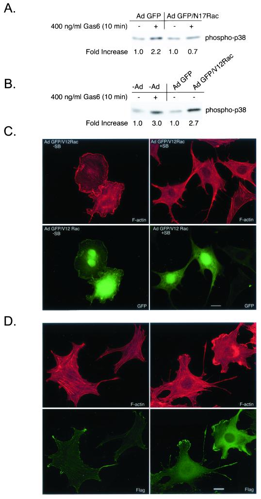
p38 activation promotes actin reorganization downstream of Rac in NLT GnRH neuronal cells. (A) NLT cells expressing Ad GFP or GFP/N17Rac were treated for 10 min with Gas6 (400 ng/ml) and then immunoblotted for phospho-p38. The data are representative of two independent experiments. The increase in phospho-p38 was calculated by setting the density of the no-Gas6 samples to 1. (B) uninfected NLT cells or those expressing Ad GFP or GFP/V12Rac were treated for 10 min with Gas6 (400 ng/ml) or vehicle and then immunoblotted for phospho-p38. The data are representative of two independent experiments. (C) NLT cells were infected with adenoviruses expressing GFP/V12Rac (see Materials and Methods), treated for 4 h with vehicle (DMSO) or SB203580 (30 μM) and then stained with rhodamine phalloidin. The data are representative of two independent experiments. Actin is shown in red and GFP is in green. Bar = 20 μm. (D) NLT cells infected with adenoviral MKK6CA and Flag-p38α were visualized with rhodamine phalloidin and anti-Flag immunocytochemistry (left panels, uninfected cells; right panels, cells infected with MKK6CA and Flag-p38α). Bar = 20 μm.

