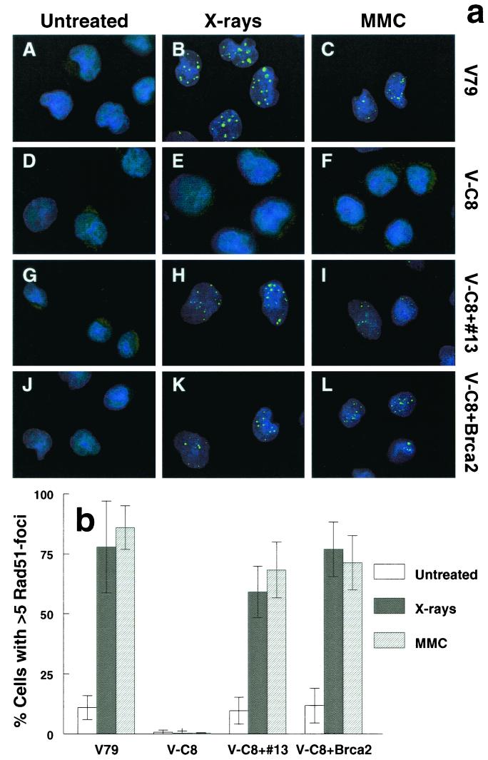
FIG. 1.
(a) Immunofluorescent visualization of Rad51 nuclear foci: wild-type V79 (A to C), V-C8 (D to F), V-C8 cells with human chromosome 13 providing the BRCA2 gene (V-C8+#13) (G to I), and V-C8 containing a BAC with the murine Brca2 gene (V-C8+Brca2) (J to L). Cells were analyzed for 8 h after treatment with either 12 Gy of X rays (B, E, H, and K) or 2.4 μg of MMC/ml for 1 h (C, F, I, and L). (b) Quantification of Rad51 focus-positive cells. A cell containing more than five distinct foci was considered positive. Each bar represents the result of scoring at least 100 cells. The error bars represent the standard error of the mean (SEM).

