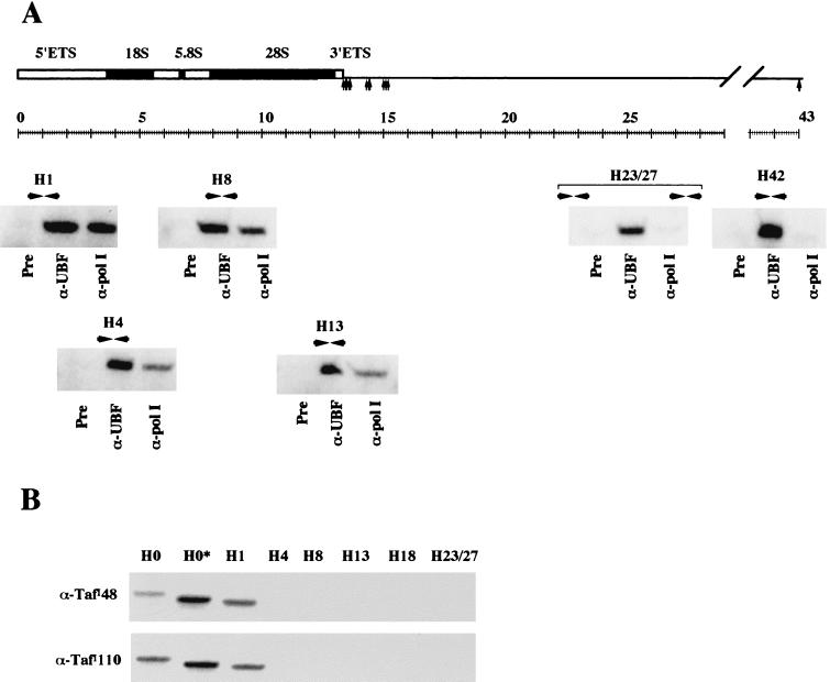
FIG. 8.
RNA Pol I and SL1 show a resticted distribution on the human rDNA repeat. (A) PCR was performed with DNA extracted from preimmune, α-UBF, and α-Pol I ChIP assays with primer pairs from across the human rDNA repeat. The locations of the primer pairs and gels of the resulting PCRs are shown in the appropriate position below a diagram of the human rDNA repeat. The source of DNA used in each PCR is shown below the gel. (B) PCR was performed with DNA extracted from α-TafI110 and α-TafI48 ChIP assays with primer pairs from across the human rDNA repeat. The identity of the primer pairs is shown above the appropriate gel lanes, and the identity of the antibody used in ChIP is shown alongside.

