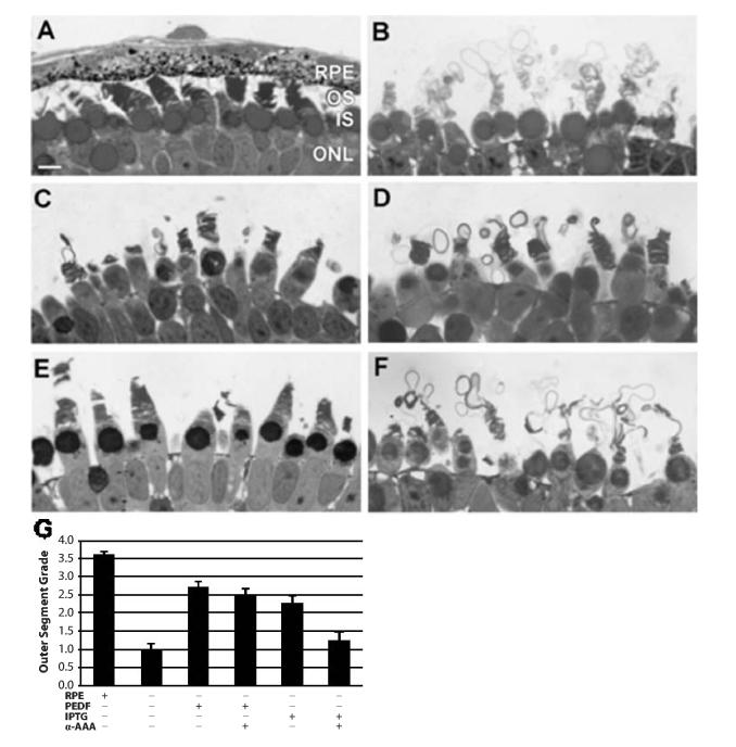
Fig. 1.

Illustrative examples of photoreceptor outer segment organization under the experimental conditions utilized in this study. (A) Control outer retina in which photoreceptors elaborated outer segments with a juxtaposed retinal pigment epithelium (RPE) layer. (B) Negative control RPE-deprived retina in which outer segments were synthesized in non-Rplemented Niu-Twitty medium. (C) RPE-deprived retina in which outer segment membranes were elaborated in medium Rplemented with 50 ng/ml purified bovine pigment epithelial-derived factor (PEDF). (D) RPE-deprived retina that was maintained in medium Rplemented with both 50 ng/ml purified bovine PEDF and 10-5 M alpha-amino adipic acid (α-AAA). (E) RPE-deprived retina that was exposed to 5 × 10⁻⁵ M isopropyl beta-D-thiogalactoside (IPTG), a permissive glycan. (F) RPE-deprived retina that was exposed to both 5 × 10⁻⁵ M IPTG and α-AAA. (G) Graphic illustration of photoreceptor outer segment grading from the various experimental conditions. RPE=retinal pigment epithelium; OS=photoreceptor outer segments; IS=photoreceptor inner segments; ONL=outer nuclear layer. Magnification bar=10 μm.
