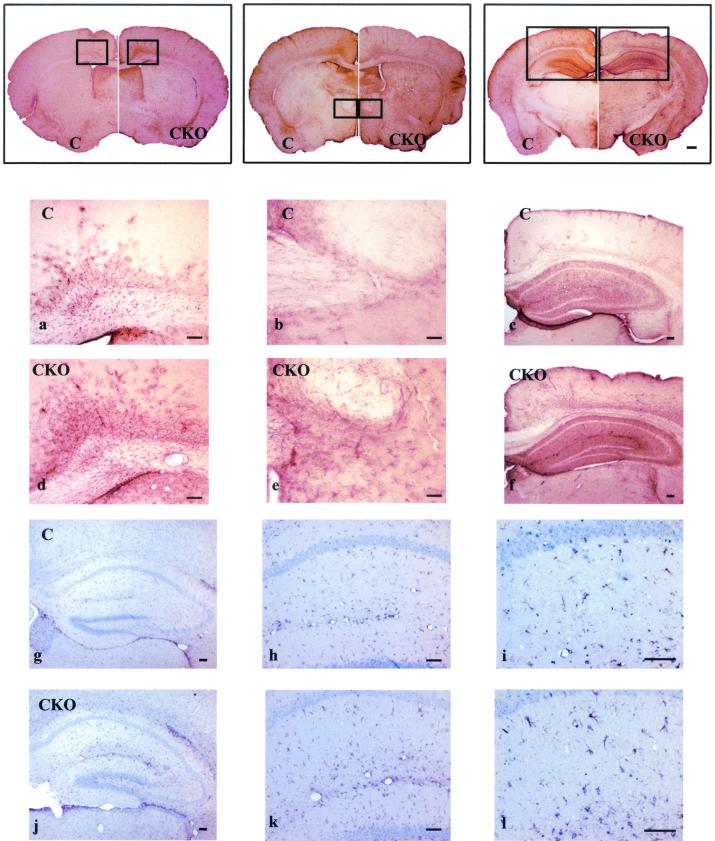
FIG. 4.
Astrocyte-specific Nf1 conditional knockout mice demonstrate moderate increases in astrocyte number. Low-power photomicrographs of GFAP immunohistochemistry of 40-μm brain sections (microtome) were aligned from rostral to caudal, with the right side of each panel representing Nf1GFAPCKO brain sections (CKO) and the left side of each panel representing brain sections from control mice (C). Nf1GFAPCKO mice demonstrate increased GFAP immunoreactivity throughout the brain. Higher magnification of the boxed areas in the brains of control mice (C) on the left side of the upper panels (a, b, and c) and of the boxed areas in the brains of Nf1GFAPCKO mice (CKO) on the right side of the upper panels (d, e, and f) revealed increased numbers of astrocytes in the corpus callosum, hippocampus, and cortex of Nf1GFAPCKO mice. Increased numbers of astrocytes without major changes in morphology were also observed in Nf1GFAPCKO hippocampal sections by GFAP and eosin staining (panels j, k, and l) compared to control mice (panels g, h, and i).

