FIG. 6.
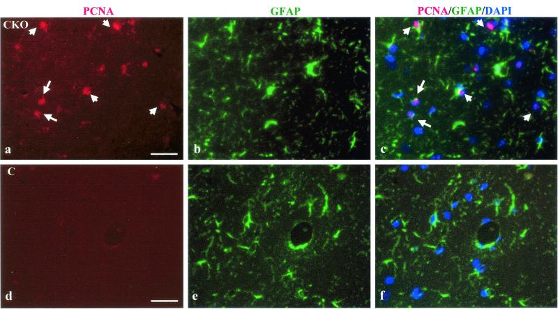
Increased astrocyte number in the brains of Nf1GFAPCKO mice is the result of increased astrocyte proliferation. PCNA immunofluorescence identifies proliferating cells (red) in the CA1 region of the hippocampus of adult Nf1GFAPCKO (CKO) mice. White arrows point to representative PCNA-labeled cells (panel a). Almost no PCNA-immunoreactive proliferating cells were present in the hippocampus of control (C) mice (panel d). GFAP immunofluorescence (green) of astrocytes in the CA1 region of the hippocampus is shown for CKO mice (panel b) and for control mice (panel e). Note that there are increased numbers of astrocytes in panel b, corresponding to the mutant mice, compared to panel e (control mice). Merged PCNA/GFAP/DAPI images are shown for CKO (panel c) and control (panel f) mice. The nuclei of the cells in these images are stained with DAPI and appear dark blue. The white arrows in the merged PCNA/GFAP/DAPI (panel c) for the Nf1GFAPCKO mice denote several double-labeled GFAP-positive astrocytes with PCNA-labeled nuclei (pink). No GFAP/PCNA/DAPI-immunolabeled cells are observed in the hippocampus of control mice (panel f).

