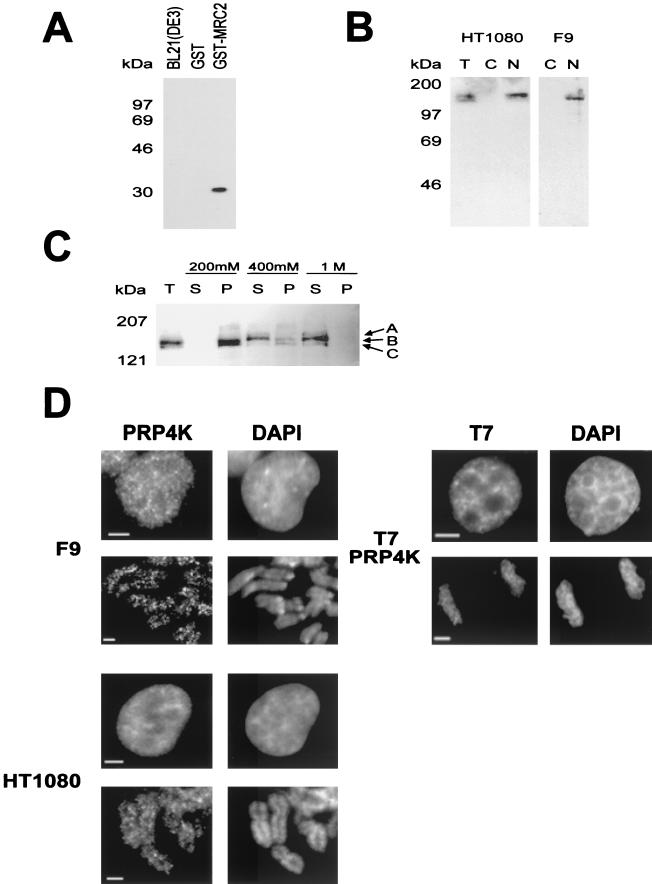
FIG. 2.
Characterization of PRP4K by Western blot analysis and indirect immunofluorescence. (A) Specificity of the anti-PRP4K antibody MRC2. Western blot analysis shows that affinity purified anti-PRP4K antibody MRC2 can detect the MRC2 epitope fused to GST (GST-MRC2) but does not cross-react with GST alone or with total bacterial protein extract. (B) Western blot analysis with the anti-PRP4K antibody MRC2 on total (T), cytoplasmic (C), or nuclear (N) extracts prepared from the human HT1080 or murine F9 embryocarcinoma cells indicates that PRP4K is a nuclear protein. (c) Salt extraction of PRP4K from human HT1080 nuclei was performed. Western blot analysis with the MRC2 antibody of soluble (S) and pellet (P) fractions of nuclei extracted with 200 mM, 400 mM, or 1 M KCl was then carried out. Total nuclei are shown for comparison (T). Three bands were detected: band A, 167 kDa; band B, 152 kDa; and band C, 147 kDa. Identical results were obtained with both the MRC1 and the H143 anti-PRP4K antibodies (data not shown). (D) Subcellular localization of PRP4K in murine F9 and human HT1080 cells by indirect immunofluorescence. Immunofluorescence of PRP4K (FITC) is shown in cells costained with DAPI to visualize the DNA. For each cell type, nuclei are shown in the first row of images, followed by mitotic chromosomes shown in the second row. In interphase cells, PRP4K is nucleoplasmic but enriched in foci that do not correspond to regions of concentrated DNA (DAPI). At mitosis, some PRP4K is also localized to mitotic chromosomes. Immunodetection of T7-tagged recombinant PRP4K (T7-PRP4K) in HT1080 cells produces a localization pattern similar to that of endogenous PRP4K in interphase cells and on mitotic chromosomes. Scale bars, 5 μm.

