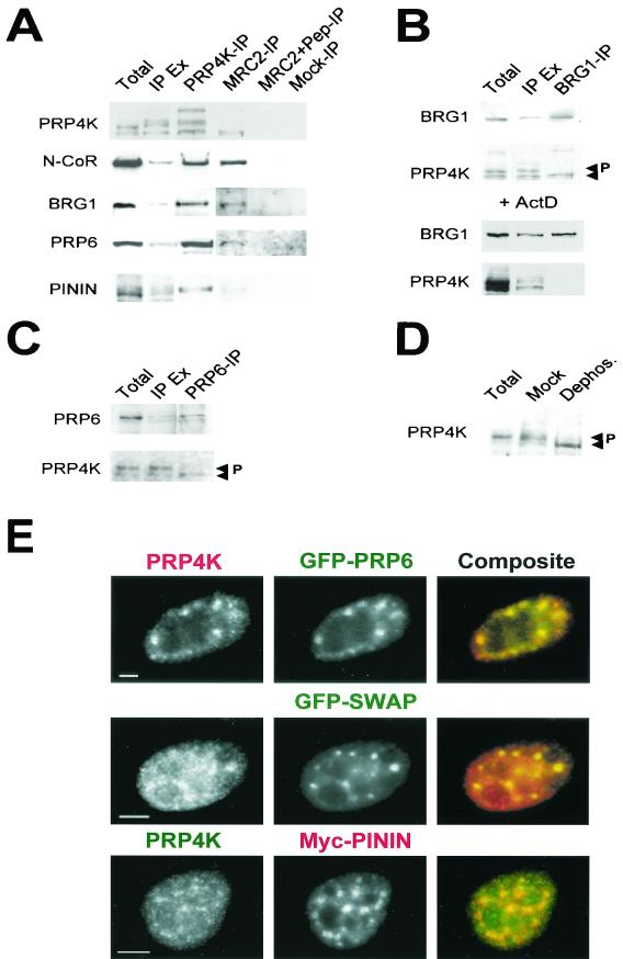
FIG. 5.
In vivo interactions of PRP4K. (A to C) Co-IP of PRP4K with pinin, N-CoR, BRG1, and PRP6. Complexes containing PRP6, BRG1, or PRP4K were immunoprecipitated from HeLa nuclear extracts with antibodies against each protein coupled to protein A- or protein G-agarose. Total, total nuclei; IP, IP of the indicated protein; IP Ex, IP extract. (A) Western blot analysis with antibodies recognizing each protein shows that N-CoR, BRG1, PRP6, and pinin coimmunoprecipitated with PRP4K by using anti-PRP4K immune serum against the MRC1 and two epitopes (PRP4K-IP) or the affinity-purified anti-PRP4K antibody MRC2 (MRC2-IP). Control IPs were carried out with MRC2 antibody preblocked with the MRC2 peptide (MRC2+Pep-IP) and with rabbit anti-sheep IgG (Mock-IP). (B) Western blot analysis of proteins immunoprecipitated with BRG1 by using anti-BRG1 antibodies (BRG1-IP). Only the fastest-migrating form of PRP4K coimmunoprecipitated with BRG1. In the presence of actinomycin D (+ActD), PRP4K fails to coimmunoprecipitate with BRG1, suggesting that the interaction between these proteins is transcription dependent. Arrows indicate two prominent forms of PRP4K; the slower-migrating form is phosphorylated (P). (C) Western blot analysis of proteins immunoprecipitated with PRP6 with anti-PRP6 antibodies (PRP6-IP). Only the fastest-migrating form of PRP4K coimmunoprecipitated with PRP6. (D) Dephosphorylation of HeLa nuclear extracts by using CIAP. Two major forms of PRP4K are indicated by the arrows; P indicates the hyperphosphorylated and slower-migrating form. Mock dephosphorylation results are shown for comparison. These results indicate that BRG1 and PRP6 interact primarily with the hypophosphorylated form of PRP4K. (E) Colocalization of PRP4K with GFP-tagged human PRP6 (GFP-PRP6), GFP-tagged murine SWAP (GFP-SWAP), or myc-tagged pinin (Myc-PININ) in HeLa cells. Endogenous PRP4K was detected with either Texas red-labeled (for GFP-PRP6 and GFP-SWAP transfections) or FITC-labeled secondary antibodies (for myc-pinin transfections). Myc-tagged pinin (Myc-PININ) was detected with Texas red-labeled secondary antibodies. Scale bars, 5 μm.

