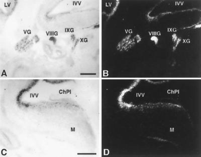
FIG. 5.
Expression of Nhlh1 in the mouse head at e13.5. Bright field (A and C) and dark field (B and D) in situ hybridizations on parasagittal sections are shown. LV, lateral ventricle; IVV, fourth ventricle; VG, trigeminal ganglion; VIIIG, vestibulocochlear ganglion; IXG, glossopharyngeal ganglion; XG, vagal ganglion; ChPl, choroid plexus; M, medulla oblongata. Scale bars represent 500 μm. No signal was observed when sections were hybridized with a sense probe (data not shown).

