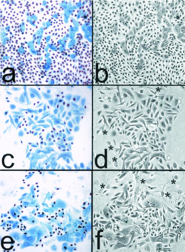
FIG. 1.
Cessation of division at the end of life span by primary human keratinocytes in culture is tightly linked to the expression and accumulation of p16INK4A. (a and b) Mid-life-span OKF4 cells serially passaged in K-sfm medium (at ≈25 PD of their ≈37-PD life span). (c and d) Late-life-span OKF4 cells serially passaged in the feeder fibroblast/FAD medium system (at ≈40 PD of their ≈45-PD life span). (e and f) Immortalized OKF4/TERT (early, feeders) cells serially passaged in the feeder fibroblast/FAD medium system (at 70 PD, which is 25 PD beyond the life span limit of the parent cell line). All three cell lines were plated at a low density (≈103 cells/cm2) and allowed to grow for 6 to 8 days before fixation. The fields shown are typical colonies that were the progeny of single cells. (a, c, and e) BUdR had been added to the medium 24 h before fixation, and cells were coimmunostained for p16 (blue) and for BUdR (red/brown). Asterisks in panels d and f indicate large, flat, p16- and BUdR-negative 3T3 fibroblast feeder cells at the periphery of keratinocyte colonies. Note that in the three cell lines shown, nearly all cells were cycling except for those containing moderate to high levels of p16. Small p16-positive cells are rare, indicating that cells become large, flat, and nondividing within several days of expressing p16. (b, d, and f) Phase-contrast images.

