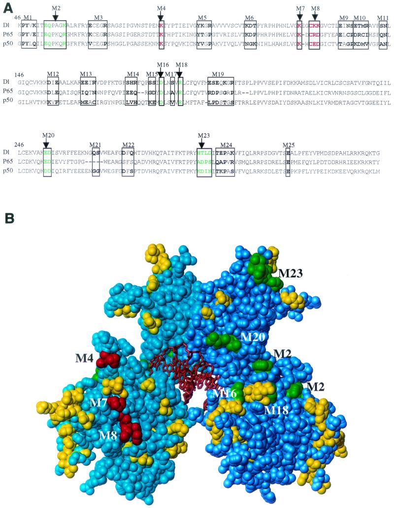
FIG. 1.
Mutagenesis of the Dorsal RHD. (A) Sequence alignment of the Dorsal RHD with those of p65 and p50. The amino acids that were mutated are indicated in bold and boxed. Amino acids changed in class I and class II mutants are colored green and red, respectively. (B) Space-filling model of the RHD. The RHD homodimer structure is based on the coordinates determined for p50 (Protein Data Bank identification no. 1NF-K). The two polypeptide chains are shown in cyan and blue, and the DNA is shown in orange. The amino acids that were altered in the mutants are shaded green (class I mutations), red (class II mutations), and yellow (phenotypically silent mutations).

