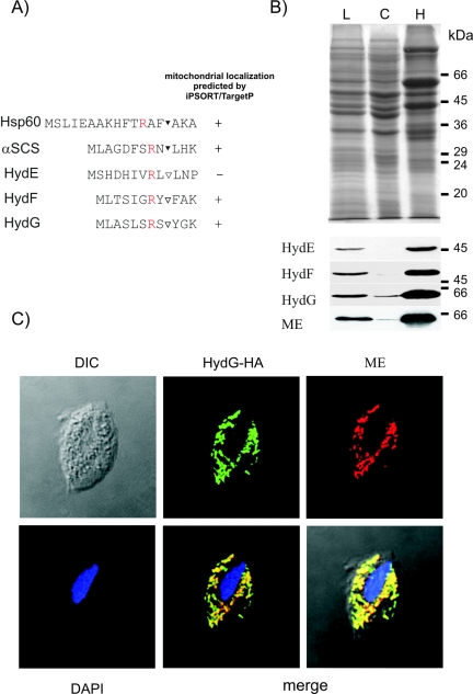
FIG. 3.
Intracellular localization of Hyd proteins in T. vaginalis. (A) Comparison of the N-terminal targeting sequences of the hydrogenosomal proteins Hsp60 and α-SCS with the N-terminal sequences of Hyd proteins. ▾, protease processing site; ▿, putative protease processing site. The typical arginine residue (R) at position −2 of the protease processing site of hydrogenosomal targeting sequences is shown in red. (B) Localization of HA-tagged HydE, HydF, and HydG overexpressed in T. vaginalis. Sodium dodecyl sulfate-polyacrylamide gel electrophoresis of HydF-overexpressing cells (top) and Western blots of cellular fractions from cells expressing tagged HydE, HydF, or HydG (bottom) are shown. ME was used as a hydrogenosomal marker protein. L, cell lysate; C, cytosolic fraction; H, hydrogenosomal fraction. (C) Immunolocalization of HydG in transfected cells shows colocalization of HA-tagged HydG with hydrogenosomal ME. DIC, differential interference contrast image; DAPI, 4′,6′-diamidino-2-phenylindole-stained image.

