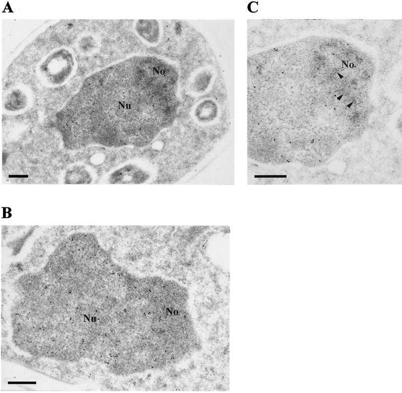
FIG. 4.
Naf1p-ZZ accumulates mainly within the nucleoplasm. Shown are electron micrographs of a whole cell (×27,600 magnification) (A), a nucleus (×41,400 magnification) (B), and a nucleolus (×46,000 magnification) (C). Naf1p-ZZ was detected by electron microscopy after treatment of the grids with anti-protein A antibodies followed by incubation with colloidal gold-conjugated protein A. No, nucleolus; Nu, nucleoplasm. In panel C, a section of the nucleolar dense fibrillar component is indicated by arrow heads. Bar = 300 nm.

