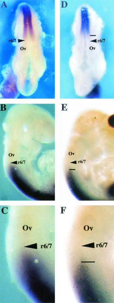
FIG. 7.
Hoxd4 gene expression in Af9 null mutant embryos. Hoxd4 expression was analyzed by whole-mount in situ hybridization (66) of wild type (A to C) and Af9−/− (D to F) E9.5 embryos. A Hoxd4 riboprobe was made from a cDNA fragment and labeled with digoxigenin (46). (A and D) Dorsal views; (B, C, E, and F) lateral views. (C and F) Magnification (×2.5) of the specimens from panels B and E. In wild-type embryos, the Hoxd4 staining extended to a boundary localized between rhombomeres r6 and r7. In the Af9−/− embryos, a posterior shift of the anterior expression limit, corresponding to approximately one rhombomere, was observed. Ov, otic vesicle.

