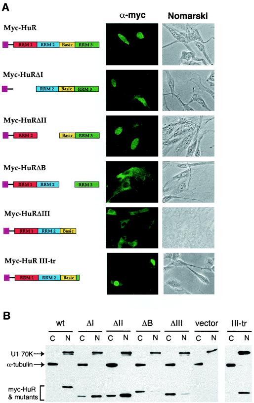
FIG. 4.
Subcellular distributions and expression levels of human HuR in mouse NIH 3T3 cells. (A) Indirect immunofluorescence microscopy study with a monoclonal antibody against the Myc epitope tag (9E10) showing the subcellular distribution of HuR and its truncated mutants (depicted by the diagram). Both phase contrast (depicted as Nomarski) and immunofluorescence (depicted as α-Myc) views are shown. (B) Western blot analysis. Cytoplasmic (C) and nuclear (N) lysates prepared from NIH 3T3 cells transfected with individual plasmids containing Myc-tagged HuR or its truncated mutant derivatives were resolved on an SDS-12% polyacrylamide gel. The blots were probed with the 9E10 monoclonal antibody for HuR and its mutant proteins, a control monoclonal antibody against α-tubulin for the cytoplasmic lysate, and a control monoclonal antibody against U1 70K for the nuclear lysate.

