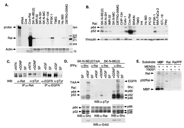
FIG. 1.
Expression pattern of Rai in neuronal cell lines and phosphorylation by activated Ret and EGFR. (A) RNase protection analysis of Rai expression in different tumor cell lines using 10 μg of total RNA and Rai (SH2 domain) (upper panel) or actin (lower panel) antisense probes. First lane, intact Rai probe; tRNA, tRNA used as a negative control; Cos-Rai, Cos cells transiently transfected with the p52Rai cDNA and used as a positive control. The specific Rai- and actin-protected fragments used are indicated above the remaining lanes. (B) Western blot analysis of Rai expression with 50 μg of whole-cell lysates from the indicated cell lines and polyclonal antibodies against the Rai CH1 region (upper panel) or vinculin for normalization (lower panel). The cell lines used in panels A and B were SK-N-BE(2), SK-N-SH, IMR-32, and CHP-134 (neuroblastomas); SK-N-MC (neuroepithelioma); PFSK-1 (primitive neuroectodermal tumor); DBTRG-05MG and T98G (glioblastomas); Hs 683 (glioma); H4 (neuroglioma); Calu-1 (lung carcinoma); Hey (ovarian carcinoma); MIA PaCa-2 (pancreatic carcinoma); GTL-16 (gastric carcinoma); and PC12 (phaeochromocytoma). Intact mouse brains (BRAIN) were also used. (C) Expression of functional Ret and EGFR in SK-N-BE(2) cells. Lysates from serum-starved (SF) cells (24 h) stimulated for 5 min with 100 ng of GDNF/ml (+GDNF), 100 ng of NTN/ml (+NTN), or 100 ng of EGF/ml (+EGF) were immunoprecipitated with anti-Ret (α-Ret) or anti-EGFR (α-EGFR) antibodies and immunoblotted with anti-Ret, anti-EGFR, and anti-pTyr (α-pTyr) antibodies, as indicated. (D) SK-N-BE(2) cells expressing endogenous Shc or Rai proteins and SK-N-BE(2)TrkA cells (engineered to express TrkA) were serum starved (24 h) (SF) and stimulated for 5 min with 100 ng of EGF/ml (+EGF), 100 ng of GDNF/ml (+GDNF), or 100 ng of NGF/ml (+NGF). Total lysates were immunoprecipitated with antibodies against the CH1 region of Shc (α-Shc) or Rai (α-Rai) and immunoblotted with antibodies against pTyr (α-pTyr) (upper panel), Shc or Rai (middle panel), or Grb2 (α-Grb2) (lower panel). Activated receptors and Grb2, Shc, and Rai polypeptides are indicated by arrows. (E) Cos cells were transiently transfected with the expression plasmids encoding Ret MEN2A, Ret Y905F, Rai, or Rai FFF. The in vitro kinase assay was performed with immunoprecipitated Ret proteins (MEN2A and Y905F) and immunoprecipitated Rai proteins (Rai and Rai FFF) as the substrates. As a positive control, we used the synthetic substrate MBP (myelin basic protein). IP, immunoprecipitate; WB, Western blot; +, present; −, absent.

