Figure 7.
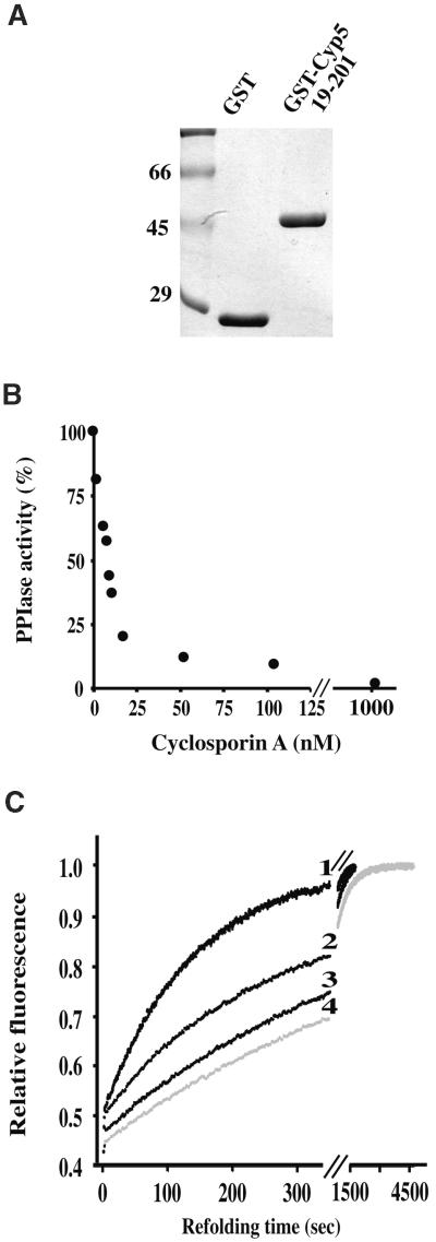
Cyclosporin A Inhibition of Cyp5 PPIase and Protein Refolding Activities.
(A) A Coomassie blue–stained SDS–polyacrylamide gel with purified GST and GST–Cyp519–201 fusion used in PPIase and protein refolding assays. Numbers indicate molecular weight markers in kilodaltons.
(B) Inhibition of Cyp5 PPIase activity by different cyclosporin A concentrations. GST–Cyp519–201 (3 nM) was preincubated with varying concentrations of cyclosporin A, and PPIase activity was measured in 35 mM Hepes buffer, pH 7.8, at 10°C.
(C) Cyp5 catalysis of slow protein refolding of RNAse T1 (0.7 μM) and inhibition by cyclosporin A. The increase of fluorescence at 320 nm is shown as a function of the time of protein refolding. Curve 1 shows 77 nM Cyp5 without cyclosporin A; curve 2, 77 nM Cyp5 with 50 nM cyclosporin A; curve 3, 77 nM Cyp5 with 100 nM cyclosporin A; and curve 4, without Cyp5 and cyclosporin A. GST did not have an effect on refolding (not shown). Measurements were performed in 35 mM Hepes buffer, pH 7.8, at 10°C.

