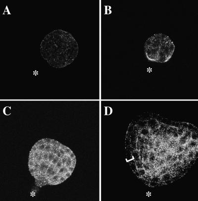
Figure 9.
Immunolocalization of Cyp5 during Embryogenesis.
Whole-mount preparations of embryos were stained with (A) preimmune serum (1:3000; control) or (B) to (D) anti-Cyp5 peptide antiserum (1:3000) followed by Cy3-conjugated secondary antibody (goat anti–rabbit; Dianova, Hamburg, Germany). Images represent internal optical sections generated by confocal microscopy. Stages of embryogenesis (Jürgens and Mayer, 1994) are shown in (A) to (D). Asterisks, basal end of embryo; bracket, epidermal cell layer.
(A) Mid-globular-stage embryo.
(B) Early-globular-stage embryo.
(C) Late globular/triangular–stage embryo.
(D) Early-heart-stage embryo. Note the low intensity of signal in the epidermal layer (bracket).

