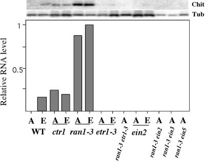
Figure 4.
The ran1-3 Mutation Results in Increased Expression of β-Chitinase.
RNA gel blot analysis of wild-type (WT), ctr1, ran1-3, etr1-3, ran1-3 etr1-3, ein2, ran1-3 ein2, ran1-3 ein3, and ran1-3 ein5 (as indicated) adult tissues grown on soil. Plants were placed in an enclosed chamber containing either air (A) or 10 μL L−1 ethylene (E) for 48 hr as indicated. Total RNA was extracted, separated by gel electrophoresis, blotted to a nylon membrane, and hybridized with either a β-chitinase probe (Chit) or a β-tubulin probe (Tub). The signals were quantified by using a PhosphorImager. The signal from the β-chitinase blot was normalized to the β-tubulin signal, and the highest ratio was then assigned a value of 1; the others were plotted relative to that. The original image of the gel blot is shown at top, and the lanes correspond to the labeling of the graph's x axis.

