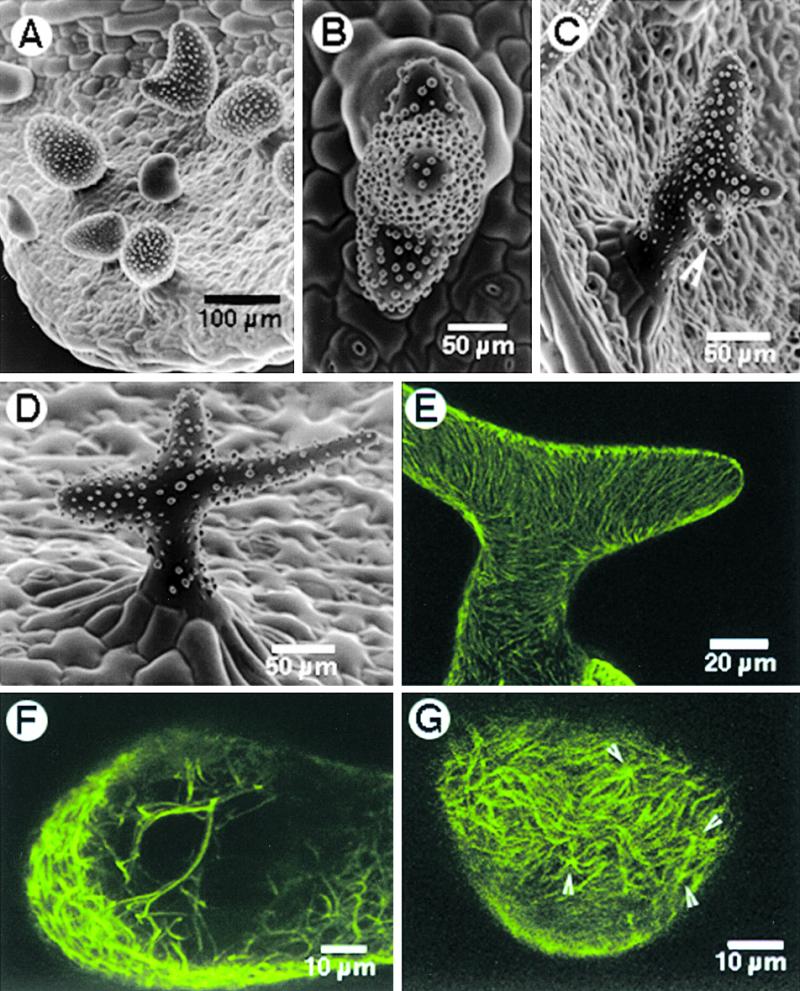
Figure 5.

Effect of Oryzalin and Paclitaxel Treatment on zwichel 9311-11 Trichomes.
(A) Treatment with 20 μM oryzalin for 2 hr resulted in radially swollen trichomes.
(B) SEM view from the top of a single trichome, showing three branch initials. The result was obtained after treating seedlings for 2 hr with 20 μM paclitaxel, followed by washing and recovery on Murashige and Skoog basal medium.
(C) A single zwichel trichome with an additional incipient branch (arrowhead). Note that the positioning of such de novo–induced branching was random.
(D) Branches extended in some of the three-branched trichomes obtained after treatment with 20 μM paclitaxel.
(E) A trichome cell with bundled cortical microtubules at 10 hr after treatment with 20 μM paclitaxel.
(F) An optical section through a trichome 10 hr after paclitaxel treatment, revealing numerous thick, subcortical, cross-linked microtubule bundles.
(G) Examination of the cortical microtubule array on a subtle bulge arising on an unbranched zwichel trichome, nearly 24 hr after the paclitaxel treatment, revealed the presence of numerous microtubule asters (arrowheads).
