
Figure 3.
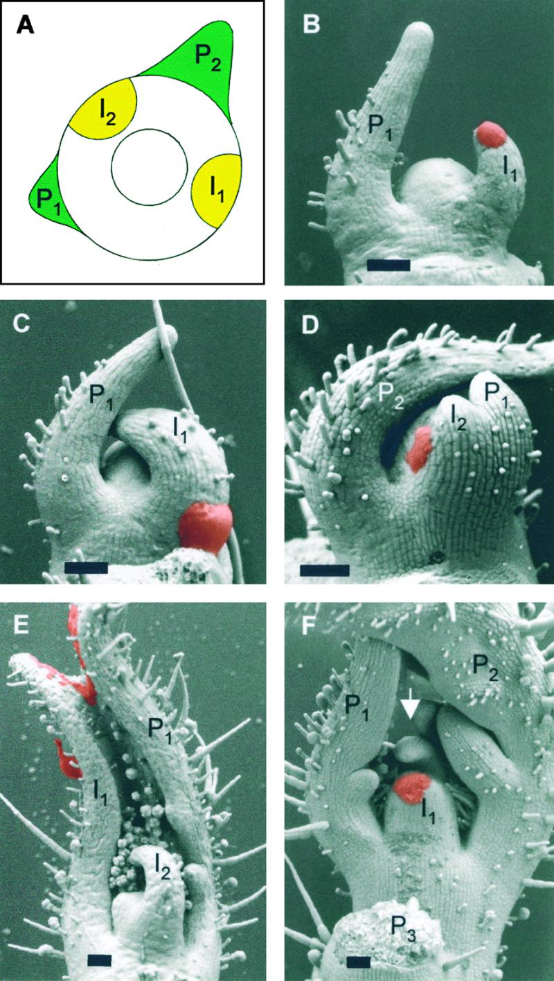
Effects of Treatments of Meristems with IAA and NPA.
(A) Schematic representation of a tomato apex with the youngest and the second youngest primordia (P1 and P2, respectively). The large circle delimits the apical meristem, and the small circle marks the central zone, which is not involved in organogenesis. The sites of incipient primordium formation (I1) and the site of the following primordium (I2) are indicated as yellow areas.
(B) Control treatment with lanolin (red paste) at the site of incipient primordium formation (I1). The primordium that formed at the I1 position has a normal size relative to P1 (cf. with Figure 1C).
(C) Increased primordium size caused by IAA treatment (red paste) at I1. P1 denotes the preexisting primordium. Note that the base of I1 is extended toward P1.
(D) Induction of an ectopic primordium by IAA (red paste) at the I2 position (between preexisting primordia P1 and P2). The ectopic primordium is fused to P1.
(E) Induction of a large fused leaf primordium caused by IAA treatment (red paste) on the entire flank of the meristem. The fused leaf includes P1 (right side) and I1 (left side). P2 (which would have been toward the viewer) was removed before scanning electron microscopic analysis.
(F) Inhibition of primordium initiation by NPA treatment at I1 (red paste) and formation of an ectopic primordium at I2 (arrow, between preexisting primordia P1 and P2).
Apices shown in (B) to (D) were examined 3 days after treatment; apices shown in (E) and (F) were analyzed 6 days after treatment.  .
.