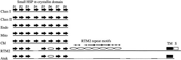
Figure 7.
Diagrammatic Representation of Known or Predicted Structural Features of Plant Small HSPs, RTM2, and Atuk.
The relative positions of the eight β strands in the small HSP α-crystallin domain are indicated by large arrows. The predicted α-helical structures in the α-crystallin–like domain of RTM2 and Atuk are shown by ovals. The repeated sequences in the central region of RTM2 are indicated by the two sets of small arrows. The region predicted to form a long α-helical structure by the Coils program (Lupas et al., 1991) is indicated by the intertwining diagram. Gaps to maintain structural alignment are indicated by the dotted lines. Chl, chloroplast; Endo, endomembrane; Mito, mitochondrial; S, serine-rich sequence; TM, predicted transmembrane sequence.

