FIG. 6.
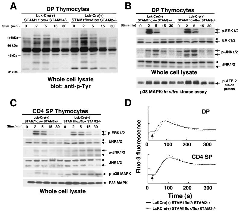
TCR-induced tyrosine phosphorylation, activation of ERK, JNK, and p38MAPKs, and intracellular calcium fluxes in thymocytes. (A and B) Double-positive thymocytes were incubated with anti-CD3 Ab for the indicated times. Induction of tyrosine phosphorylations of total proteins (A), ERK (B) and JNK (B) in whole-cell lysates was analyzed by immunoblotting. p38MAPK activity was measured using an immunocomplex kinase assay (B). (C) CD4 single-positive thymocytes were incubated with anti-CD3 Ab for the indicated times. Induction of ERK, JNK, and p38MAPK phosphorylation was analyzed by immunoblotting. (D) Intracellular calcium fluxes were measured in double-positive thymocytes or CD4 single-positive thymocytes using Fluo-3. Biotinylated anti-CD3 Ab was first added at 0 s, and then streptavidin was added at 20 s (arrow indicated) for cross-linking.

