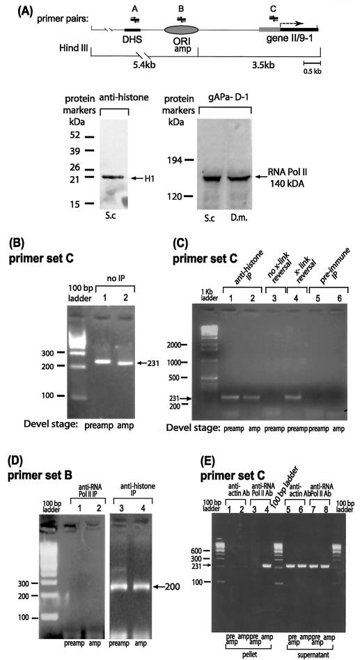
FIG. 6.
ChIP reveals in vivo occupancy by RNA polymerase II near the right-hand border of the initiation zone for DNA amplification. (A) Map of the region of interest in locus II/9A of Sciara, with PCR primer sets A to C indicated. Other details of the map are the same as in Fig. 2. Sites cleaved by HindIII are shown below the map. Western blots are shown for reaction of protein from S. coprophila (S.c.) or D. melanogaster (D.m.) larval homogenate with antibody against the 140-kDa subunit of RNA polymerase II (gAPα-D1) or histones. Only histone H1 is detected in 0.5 M NaCl extracted material. (B to E) Gel electrophoresis of ethidium bromide-stained PCR products with primer set C (panels B, C, and E) or primer set B (panel D) after ChIP with the antibody shown or after the indicated control treatments.

