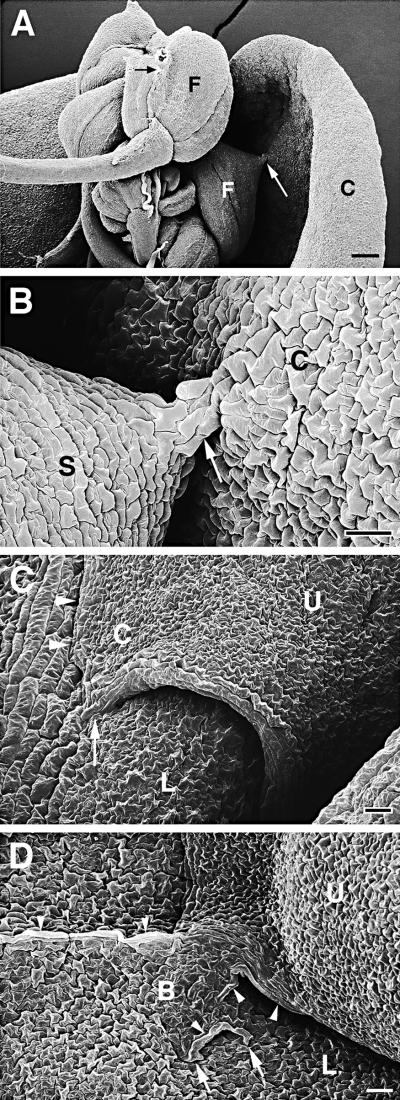
Figure 5.
Scanning Electron Microscopy of Organ Fusions of Cutinase-Expressing Arabidopsis Plants.
(A) Five flowers with fused flower organs were fused to each other, and one flower was fused to a cauline leaf. Cell contacts between two sepals of one flower (small black arrow) and between a sepal and the cauline leaf (large white arrow) were visible. C, cauline leaf; F, flower.  .
.
(B) Enlargement of (A). Small cell bridge (arrow) between sepal and cauline leaf. Epidermal cells appeared slightly larger in the cell bridge than in the unfused tissue. C, cauline leaf; S, sepal.  .
.
(C) Two overlapping rosette leaves were fused to each other. The normally oval upper leaf formed a corner in the area of fusion. Suture with hidden cell contacts is indicated with arrowheads; visible cell contacts are indicated with an arrow. C, leaf corner; L, lower leaf; U, upper leaf.  .
.
(D) A large cell bridge formed between two fused leaves. Large parts of the suture were not visible. Several cell contacts are visible (arrows). In part of the cell bridge, large epidermal cells developed (arrowheads), whereas the epidermal cell layer of other areas was normally differentiated. B, cell bridge; L, lower leaf; U, upper leaf.  .
.

