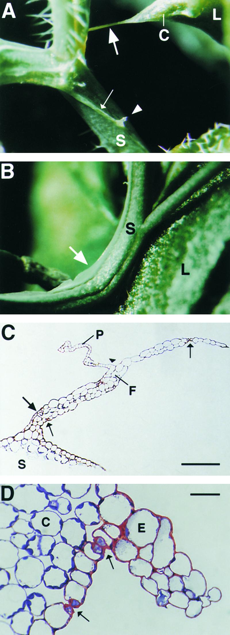
Figure 6.

Unusual Epidermal Protrusions Formed in Cutinase-Expressing Arabidopsis.
(A) A pointed corner with a long epidermal attachment filament (large arrow) developed on a leaf after organ fusion with a stem under high mechanical stress. Epidermal ridges on a distorted stem are indicated by a small arrow; a protrusion originating from the ridge is marked by an arrowhead. C, leaf corner; L, leaf; S, stem.
(B) Epidermal protrusion in a bent stem segment (arrow). L, leaf; S, stem.
(C) Semithin section through an attachment filament. The large arrow indicates an area of high variability in size of epidermal cells; the small arrows indicate the stomata; and the arrowhead indicates the periclinal division plane. F, filament; P, protrusion; S, stem. Bar = 50 μm.
(D) Semithin section through a ridge protruding from a distorted stem segment. Arrows indicate the stomata. C, cortical cell; E, epidermal cell.  .
.
