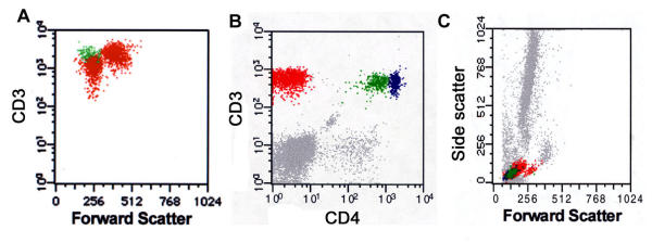
Figure 3.
Mycosis fungoides in peripheral blood with multiple aberrant T-cell populations. A. CD3 versus forward scatter identifies two abnormal populations, one large and one small, that are distinct from normal T cells. Examination of PB smear identified small (cerebriform) and large (transformed) tumor cell populations (not shown). B. Another case of MF in PB shows two atypical T-cell population; one CD4dim(+) (colored green) and another CD4(-) (red) distinct from the normal CD4(+) T-cells (blue). The CD4(-) population was also negative for CD8 (data not shown). C. Forward/side scatter histograms revealed the CD4(-) population predominantly comprised the larger (transformed) tumor lymphocyte population that was noted on PB smear.

