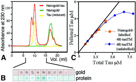
Fig. 5. (A) Separation of free nanogold from labelled protein by gel filtration (red curve). (B) Fractions of the first peak were dotted onto nitrocellulose paper and stained to detect the presence of gold and protein. The second peak (free gold without protein) did not bind to the paper. (C) Binding assays of labelled and unlabelled triple mutant tau (4R-tauTM). The curve shown is for assembly with GMPCPP; results for GTP were similar.

An official website of the United States government
Here's how you know
Official websites use .gov
A
.gov website belongs to an official
government organization in the United States.
Secure .gov websites use HTTPS
A lock (
) or https:// means you've safely
connected to the .gov website. Share sensitive
information only on official, secure websites.
