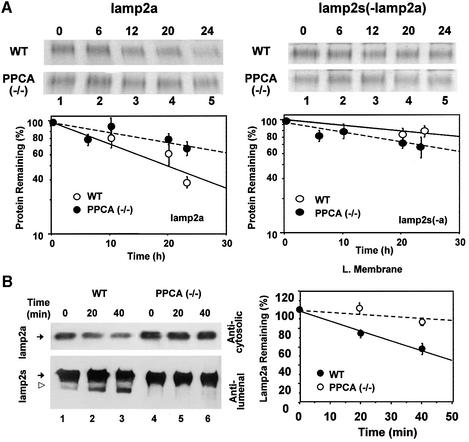
Fig. 5. Impaired lamp2a degradation in PPCA-deficient fibroblasts. (A) Autofluorography of the sequential immunoprecipitation of lamp2a first (left) and all the remaining isoforms of lamp2 second (right) from metabolically labeled wild-type and PPCA(–/–) mouse skin fibroblasts. Values are means ± SE of three different experiments similar to the ones shown in the upper panels. (B) Immunoblot analysis for lamp2a (top) and all lamp2 isoforms (bottom) in lysosomal membranes from wild-type (WT) or PPCA(–/–) mouse skin fibroblasts at different times after incubation at 37°C. The open arrowhead indicates the truncated form of lamp2 lacking the cytosolic/transmembrane region (Cuervo and Dice, 2000b). Right shows the densitometric quantification (mean ± SE) of four different experiments similar to the one shown here. Values are expressed as percentage of remaining lamp2a, and are means ± 1 SE of three different experiments.

An official website of the United States government
Here's how you know
Official websites use .gov
A
.gov website belongs to an official
government organization in the United States.
Secure .gov websites use HTTPS
A lock (
) or https:// means you've safely
connected to the .gov website. Share sensitive
information only on official, secure websites.
