Figure 5.
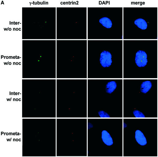
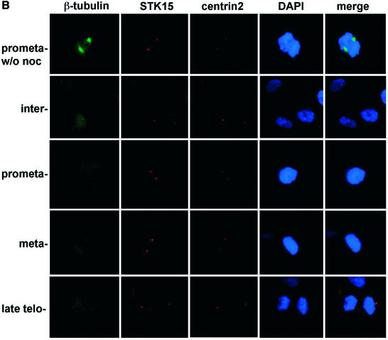
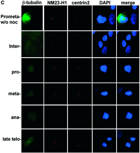
(Previous two pages and above) The centrosome localization of STK15 and NM23-H1 is not microtubule dependent. (A) IMR90 cells grown on coverslips were treated with or without nocodazole and fixed for immunofluorescence. The centrosome was visualized by staining with anti-centrin2 and anti-γ-tubulin antibodies. As predicted, the signal representing centrosome-associated γ-tubulin becomes significantly weakened, especially in mitotic cells (prometaphase cells here), upon nocodazole treatment (labeled w/ noc on the left), as compared to that seen in cells without nocodazole (labeled w/o noc on the left). (B) STK15 and (C) NM23-H1 still localize to centrosome in nocodazole-treated cells throughout the cell cycle. IMR90 cells treated with or without nocodazole (w/o noc) were fixed and triple labeled for β-tubulin (FITC), STK15/NM23-H1 (Cy5), centrin2 (Texas red) and DNA (DAPI). The nocodazole treatment totally dissipates the microtubule spindles so the cells with nocodazole fail to show the characteristic β-tubulin staining pattern (the β-tubulin columns). Cell cycle stages are indicated on the left.


