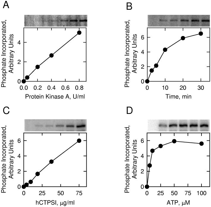
Fig. 7.
Phosphorylation of E. coli-expressed human CTP synthetase 1 by protein kinase A. Panel A, pure human CTP synthetase 1 (1 μg) was incubated with [γ-32P]ATP (50 μM) and the indicated amounts of protein kinase A for 10 min. Panel B, pure human CTP synthetase 1 (1 μg) was incubated with protein kinase A (1 U/ml) and [γ-32P]ATP (50 μM) for the indicated time intervals. Panel C, protein kinase A (1 U/ml) and [γ-32P]ATP (50 μM) were incubated with the indicated concentrations of pure human CTP synthetase 1 for 10 min. Panel D, protein kinase A (1 U/ml) and pure human CTP synthetase 1 (1 μg) were incubated with the indicated concentrations of [γ-32P]ATP for 10 min. Following the phosphorylation reactions, samples were subjected to SDS-PAGE. The SDS polyacrylamide gels were dried and the phosphorylated proteins were subjected to phosphorimaging analysis. The relative amounts of phosphate incorporated into human CTP synthetase 1 were quantified using ImageQuant software. Portions of the images with the phosphorylated human CTP synthetase 1 are shown above each graph. The data are representative of two independent experiments.

