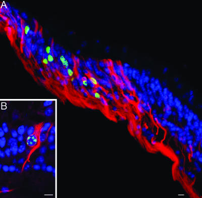
Fig. 3.
Proliferating cells are neuroprogenitors. (A) Sections of the retinal periphery were stained with antibodies to nestin (red) and BrdU (green), and nuclei were counterstained with DAPI (blue). Numerous cells contained nestin-positive bundles of cytoskeletal filaments and exhibited the elongate shape typical of embryonic neuroepithelial precursors. In many of these cells, the nucleus was BrdU-positive. (B) At higher magnification, BrdU-stained nucleus and nestin-positive cytoplasmic filaments were colocalized in the same cell. (Scale bars: 10 μm.)

