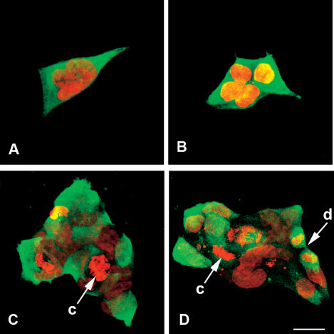
Fig 1.
Immunostaining of E11′L cells. E11′L cells immunostained with antibody to p26 and Alexa Fluor®488-labeled anti-rabbit IgG secondary antibody (green) were incubated with propidium iodide to reveal nuclei (red) and examined by confocal microscopy. c, chromosomes; d, dividing cell. Bar = 25 μm (D). All figures are the same magnification

