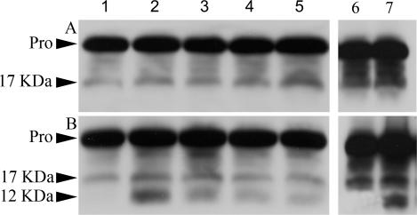
Fig 7.
Inhibition of procaspase-3 activation by p26. E11′L cells (A) and cells transfected with vector only (B) were either heat shocked at 46°C or exposed to staurosporine for 6 hours, as described in Materials and Methods. Protein extracts were prepared from these cells, electrophoresed in 12.5% SDS polyacrylamide gels, blotted to nitrocellulose, and probed with anti–caspase-3 antibody. Lanes 1– 5: protein extracts from cells heat shocked for 0, 15, 30, 45, and 60 minutes, respectively. Lanes 6, 7: extracts from cells exposed to 0.0 and 0.2 μm, respectively, of staurosporine for 6 hours. Approximately equal amounts of protein were applied to each lane. Pro, procaspase-3; 17 kDa, 17-kDa fragment from procaspase-3; 12 kDa, 12-kDa fragment from procaspase-3

