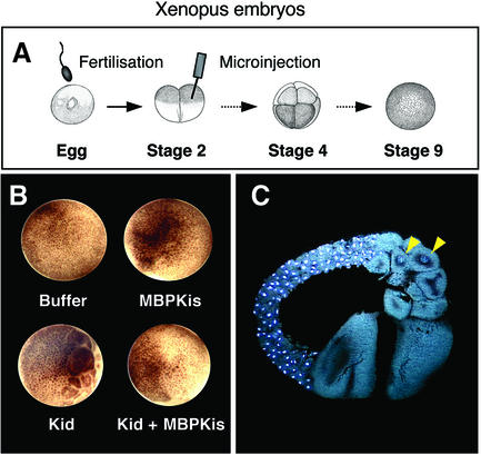
Fig. 3. Kid inhibits cell proliferation in X.laevis embryos; Kis neutralizes this effect. (A) Scheme depicting microinjection experiments of Xenopus embryos. One blastomere of a two-cell embryo was injected with either Kid, MBPKis, Kid plus MBPKis or buffer. Development was allowed to proceed until the control uninjected embryos reached stage 9 (late blastula). (B) Representative embryos from the experiment shown in (A) when buffer, MBPKis, Kid, or Kid plus Kis proteins were microinjected. Almost identical results were obtained with all the embryos injected with Kid (43 in total), MBPKis (15 in total) and Kid plus MBPKis (19 in total). (C) A section of one embryo microinjected into one blastomere with Kid protein. Embryos were fixed, paraffin-embedded, sectioned and stained for DNA. The uninjected half embryo developed normally whereas the Kid-injected half showed very few cells, most of which were anucleate. The yellow arrowheads indicate the only two nuclei present in the Kid-injected half embryo.

An official website of the United States government
Here's how you know
Official websites use .gov
A
.gov website belongs to an official
government organization in the United States.
Secure .gov websites use HTTPS
A lock (
) or https:// means you've safely
connected to the .gov website. Share sensitive
information only on official, secure websites.
