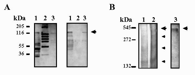
Fig. 3.

SDS-PAGE (left) and immunoblotting (right) of purified β-Galactosidase from K. lactis (A). β-Galactosidase is indicated by an arrow. Western-blot was incubated with mouse immune serum. Lane 1: 75 μg of a crude extract from K. lactis; lane 2: Molecular weight markers; lane 3: 5 μg of β- galactosidase from K. lactis purified by affinity chromatography. (B) Electrophoresis on a non-denaturing gradient gel (5-15%) stained with ONPG and later Coomassie brilliant blue G250 (left) and with BNG (right). Bands with β-Galactosidase activity are indicated by higher arrows. Different forms of β-Galactosidase are shown by smaller arrows. Different concentrations of b-Galactosidase from Maxilact LX-5000 were loaded. Lane 1:20 μg; lane 2:40 μg; lane 3:80 μg.
