
Figure 1.
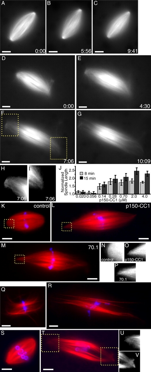
Dynein/dynactin inhibition increases the length of spindle microtubules in the presence or absence of centrosomes. (A–C) Tubulin distribution in untreated spindles during live recordings. (D–G) p150-CC1 addition (2 μM, ∼3 min before image at t = 0) caused spindles to increase in length. (H and I) Higher magnified spindle pole regions indicated in F (Videos 1 and 2). (J) p150-CC1 was added to assembled spindles, samples were fixed after 8 or 15 min, spindle lengths were measured (mean ± SD, n = 15, two independent experiments), and normalized to the length of untreated spindles (40 μm). (K–M) Spindles fixed 8 min after addition of control buffer (K), 2 μM p150-CC1 (L), or 1 mg/ml 70.1 (M) (tubulin, red; DNA, blue). (N–P) Higher magnified, contrast-adjusted regions indicated in K–M, respectively. (Q and R) Spindles assembled in 18 μm p50 dynamitin were treated with control buffer (Q) or 2 μm p150-CC1 (R) and fixed after 15 min (tubulin, red; DNA, blue). (S and T) Spindles assembled in the absence of centrosomes, around DNA-beads (tubulin, red; DNA, blue). (S) Buffer control. (T) p150-CC1–treated (2 μM, 8 min). (U and V) Higher magnified, contrast-adjusted regions indicated in R. Times are in min:s. Bars, 10 μm.