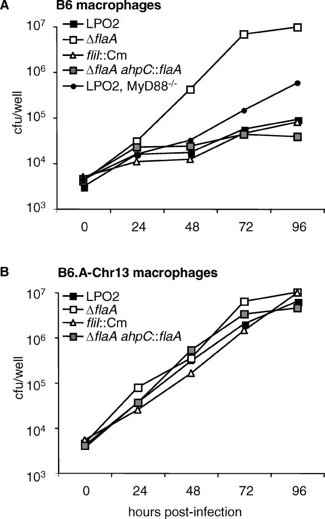
Figure 2. Growth of Legionella in Bone Marrow–Derived Macrophages.
Growth of Legionella strains in bone marrow–derived macrophages from (A) B6 mice or (B) B6.A-Chr13 mice (which carry the permissive A/J allele of Naip5). A confluent layer of macrophages were grown in 24-well plates and were infected with the indicated Legionella strains at an MOI of 0.02. Growth of wild-type Legionella (LP02) in B6-backcrossed MyD88−/− macrophages is also depicted in (A). After addition of bacteria, the plate was spun at 400 g for 10 min. One hour after infection, the media was changed. At daily intervals (starting with 1 h post-infection), a timepoint was taken. Macrophages were lysed with sterile water and the lysate combined with supernatant from the same well. Colony-forming units per well were determined by plating dilutions onto BCYE plates.

