
Figure 2.
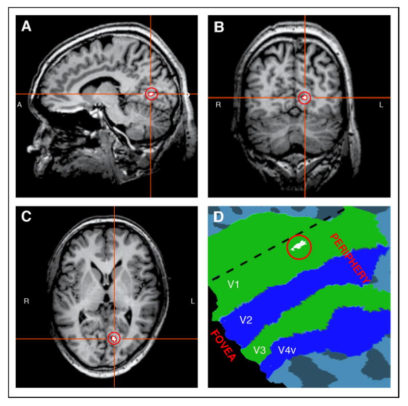
Cortical representation of the blind spot in V1, plotted in white and highlighted by a surrounding red circle, shown in sagittal (A), coronal (B), and transverse (C) views for a representative subject’s brain. This region responds more to stimulation of the fellow eye than to stimulation of the blind-spot eye. The V1 representation of the right eye’s blind spot is located in the fundus of the calcarine sulcus in the left hemisphere. D: V1 blind-spot region plotted on a flattened cortical map of retinotopic visual areas, V1- V4. The representation of the blind spot is located near the horizontal meridian (dashed line) in the periphery of V1.